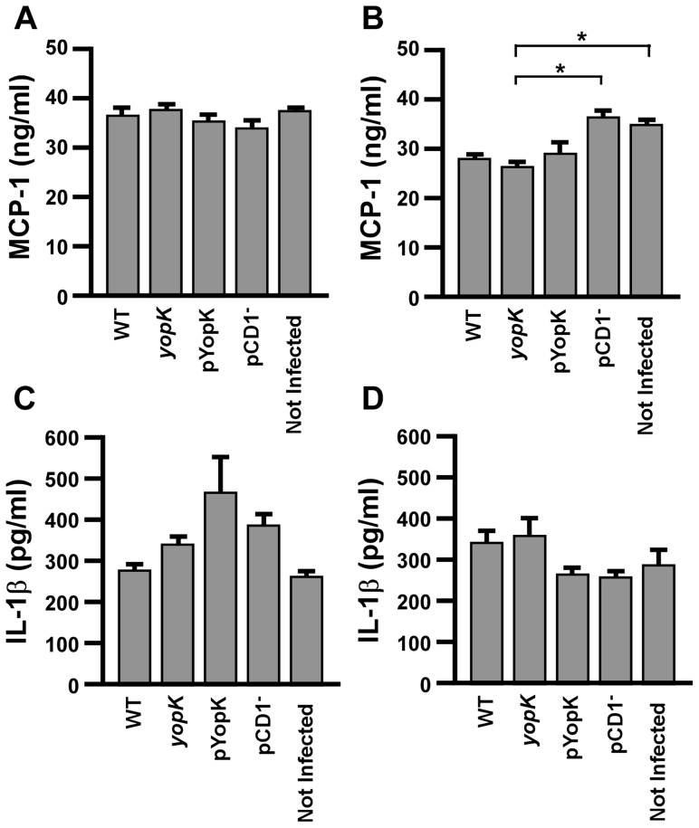
Figure 8. YopK does not directly impact secretion of pro-inflammatory cytokines from macrophages.
RAW 264.7 macrophages (A, C) and MH-S alveolar macrophages (B, D) were plated at a concentration of 1×10⁶ cells per well. Cells were activated with IFN-γ 4 hours prior to infection by the indicated strains of Y. pestis KIM D27 or CO92 pCD1− at a multiplicity of infection of 20, or were left not infected. After 8 hours of infection, supernatants were analyzed for MCP-1 (A–B) and IL-1β (C–D) by ELISA. *p<0.05, analyzed by one-way ANOVA followed by Tukey post-hoc test. Data shown are representative and collected from two independent experiments with duplicate wells per sample.

