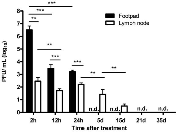
Figure 2. Mycobacteriophage D29 dissemination in footpads and DLN of mycobacteriophage D29-treated mice.

Mice were infected subcutaneously in the left footpad with 5.5 log10 AFB of M. ulcerans strain 1615. After the emergence of macroscopic lesion (33 days post infection; footpad swelling of 3.0 mm) mice were subjected to treatment with a single dose of subcutaneous injection of mycobacteriophage D29. Phage titres were assessed by plaque forming units. n.d., not detected. Results are from one representative experiment of two independent experiments. The bars represent the mean ± SD (n = 5). Significant differences were performed using Student's t test (**, p≤0.01, ***, p≤0.001).
