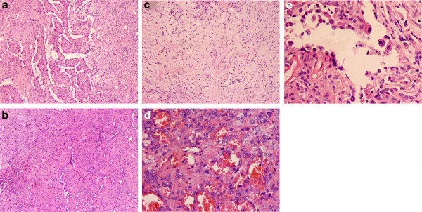
Figure 2.

Four major histologic patterns of pulmonary SH by hematoxylin-eosin stains. Pulmonary SH showed papillary (a, ×100), solid (b, ×100), sclerotic pattern (c, ×100), and hemorrhagic (d, ×400). (e) Lining cuboidal cells and stromal round cells (×400).
