Figure 1.
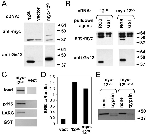
Effector binding and conformational activation of myc-tagged, constitutively activated Gα12. Molecular weight markers (in kDa) are indicated at right of panels where applicable. All results shown are representative of two or more independent experiments. (A) Expression and solubilization of Gα12QL (12QL) and myc-tagged Gα12QL (myc-12QL) transiently expressed in HEK293 cells. Cells transfected with the vector pcDNA3.1 are included as a negative control (vector). Detergent-soluble extracts were prepared by high-speed centrifugation and subjected to SDS-PAGE and immunoblotting, using either anti-myc (Zymed) or anti-Gα12 (Santa Cruz Biotechnology) antibodies as described in Methods. (B) In vitro binding of myc-tagged and untagged Gα12QL by p115RhoGEF. HEK293 cells extracts containing myc-Gα12QL were subjected to protein interaction assays (see Methods) using an immobilized GST fusion of the RH domain of p115RhoGEF (RGS) or GST alone (GST). Samples were washed, separated by SDS-PAGE, and analyzed by immunoblotting using antibodies described above. (C) Specificity of myc-Gα12QL detection in interaction assays. HEK293 cells transfected with either myc-Gα12QL (myc-12QL) or the empty pcDNA3.1 plasmid (vect) were lysed and assayed for binding to GST fusions of the RH domain of p115RhoGEF (p115) or LARG, or GST alone (GST). Immunoblot analysis was performed using anti-Gα12 antibody as described above. (D) Serum response element (SRE) luciferase activation by myc-Gα12QL. HEK293 cells grown in 12-well plates were co-transfected with the plasmids SRE-L (0.2 μg) and pRL-TK (0.02 μg), plus 0.1 μg of the plasmid indicated on the X-axis. Y-axis values show firefly luciferase signal normalized for Renilla luciferase signal within each sample. (E) Trypsin protection assays of myc-tagged Gα12. Lysates from HEK293 cells transfected with myc-Gα12QL (myc-12QL) or the constitutively GDP-bound Gly228Ala mutant of wildtype Gα12 (myc-12G228A) were subjected to trypsin digests as described in Methods. Immunoblot analysis was performed using J169 antibody [25] at 1:700 dilution.

