
Figure 2. Proportion of Ae. aegypti with a detectable infection after being held at high temperatures.
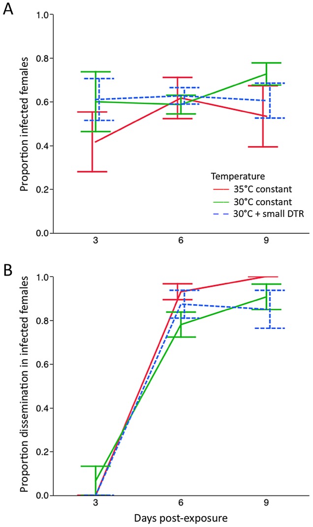
Females were held at 30°C and 35°C constant, and 30°C with a small DTR and sampled at days 3, 6 and 9 post-exposure to an infectious DENV-1 blood meal. A) Body infection, representing a detectable infection of the midgut tissue. B) Levels of infection in head tissue, representing a detectable disseminated infection.