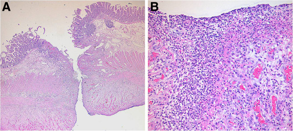
Figure 1.

Histopathology showing nonspecific inflammation and excluding IBD, vasculitis, and vascular thrombi. The low power view (A) reveals an ulcer with perforation (H&E, x20). The high power view (B) reveals necrotic debris intermixed with inflammatory cells (predominantly neutrophils) and granulation tissue formation at the perforated ileal wall (H&E, x200).
