
Figure 1.
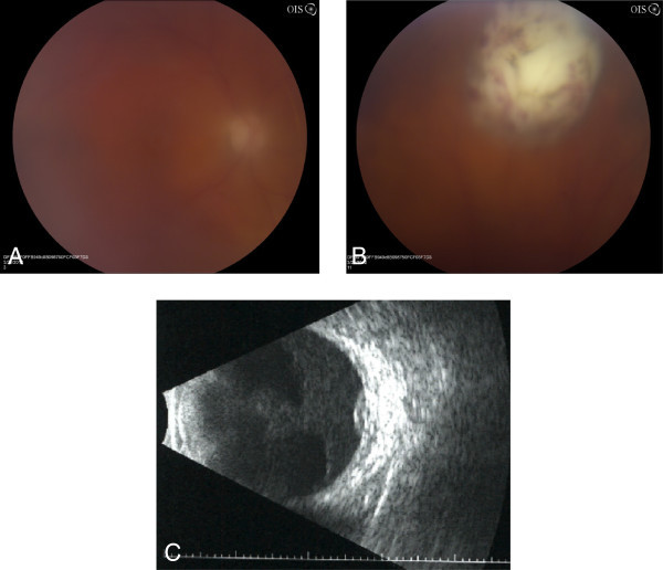
Photograph and B-scan ultrasound of the left eye. (A) Fundus photograph of the left eye at presentation showing 2+ vitreous haze obscuring the posterior pole. (B) At the 12:30 meridian, there is a dome-shaped region of retinal and subretinal whitening with overlying punctate retinal hemorrhage. (C) B-scan ultrasound confirming the presence of a superior subretinal mass measuring 1.7 mm in height and 7.1 × 7.2 mm in basal diameter.