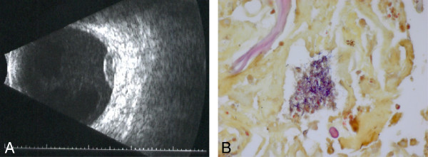
Figure 2.

Repeat B-scan ultrasound and pathologic evaluation. (A) Repeat B-scan ultrasound 1 week following initial presentation confirmed enlargement of the lesion measuring 2.6 mm in height and 9.7 × 10.3 mm in basal diameter. (B) Pathologic evaluation of the subretinal aspirate and vitreous specimen showed a cluster of gram-positive filamentous organisms consistent with Nocardia species (Brown and Brenn method, ×100).
