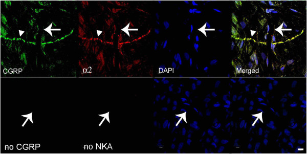
Figure 1.

Fluorescence images of CGRP and Na,K-ATPase alpha-2 in meninges. In the top row, CGRP is labeled with fluorescein on the left, Na,K-ATPase alpha-2 is visualized with Texas red, nuclei are DAPI-stained, and the merged image is on the right. The large arrow indicates a capillary and the smaller arrowhead indicates a trigeminal nerve. The lower row images are negative controls when no primary antibody was used. The notable finding is that Na,K-ATPase alpha-2 is expressed at the capillary and nerve fiber. Scale bar = 10 μm.
