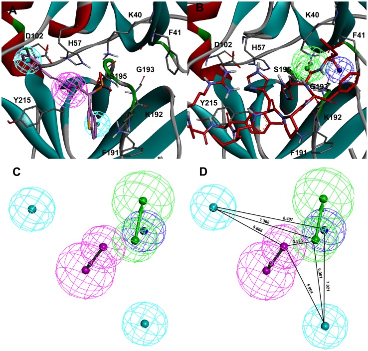
Figure 10. Development of hybrid pharmacophore model.
The key pharmacophoric features generated from the binding modes of (A) C1 and (B) Ang I. The amino acid residues of the enzyme are shown in gray thin stick form whereas the bound C1 and Ang I are shown in thick stick form. The secondary structure cartoon of the enzyme is colored based on the hydrophobicity of the amino acid residues. (C) Final hybrid pharmacophore model (D) Hybrid pharmacophore model with distance constraints.

