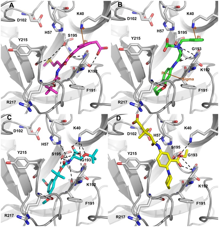
Figure 13. Molecular docking results.
The binding modes of (A) Hit 1 (B) Hit 2 (C) Hit 3 and (D) Hit 4 at the active site of the enzyme. The amino acid residues and bound ligands are shown in stick representations. The hydrogen bond, and π-interactions are shown in black dashed, and brown solid lines. Only polar hydrogen atoms are added for clarity.

