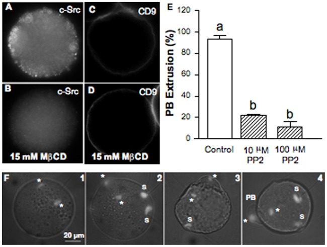
Figure 8. Effect of cholesterol depletion on c-Src and CD9 subcellular localization. Src-family kinase role on second polar body extrusion.
(A) Cortex localization of the raft-associated tyrosine kinase c-Srcassessed by indirect immunofluorescence. (B) Cytoplasmic relocation of the c-Src kinase after MβCD treatment. No primary antibody controls were negative. Staining of a total of 14 control oocytes and 6 MβCD-treated oocytes. Within each group, oocytes showed the same staining pattern. (C) Plasma membrane localization of the CD9 tetraspanin, a non-raft protein. (D) CD9 remained at the plasma membrane after MβCD treatment. Staining of a total of 18 control oocytes and 18 MβCD-treated oocytes. In both groups, oocytes showed the same staining pattern. (E) Effect of Src-family kinase inhibition assessed by incubation of oocytes with PP2 on the extrusion of the second polar body (PB). Data represent the mean ± SEM of 3 independent experiments from a total of 77 control oocytes and 85 or 69 oocytes treated with PP2 at 10 or 100 µM, respectively. (F) DAPI-stained images illustrating 1- a blocked telophase, 2- the beginning of the formation of the PB, 3- its almost complete formation, and 4- an extruded PB. Comparison of mean values was performed using Bonferroni test. Different letters (a-b) denote significant differences (P<0.05). *: oocyte chromatin; S: sperm decondensed chromatin; PB: Polar Body.

