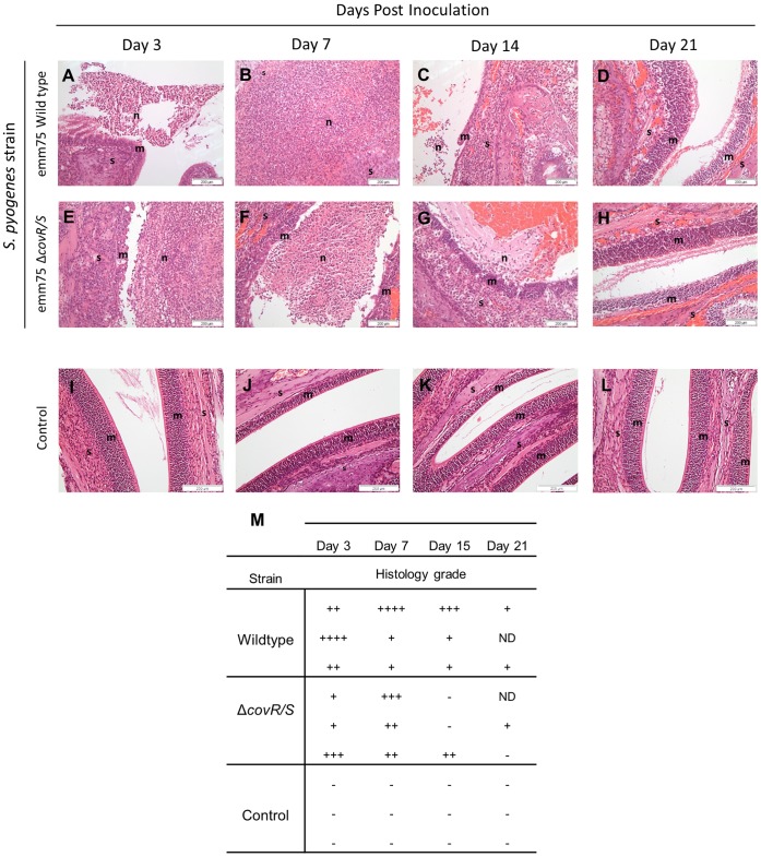
Figure 8. Histopathological analysis of the caudal nasal cavity during long term nasal infection.
Photomicrographs demonstrating five week old female FVB/n mice infected with either the emm75 S. pyogenes (A–D) or the emm75 ΔcovR/S strain (E–H) (1.5×10⁸ cfu per dose, n = 3 per group) were taken at days 3, 7, 14 and 21 after inoculation (Haemotoxylin & Eosin staining). n = Neutrophilic Exudate, m = Nasal mucosa, s = Nasal stroma. Scale bar as shown. Damage to the nasal mucosa with surface neutrophilic exudate was apparent at day 3 post inoculation with both strains (A & E). The nasal epithelia of mice in both groups were widely eroded or ulcerated by day 7 (B) than those infected with the ΔcovR/S strain (F). At day 14 the inflammation had begun to resolve in both strains (C & G). By day 21, mice in all groups had histologically normal mucosa (D & H). Control mice over the time course are shown in (I–L). Semi quantitative analysis of the histopathology was undertaken to determine the severity of infection and assigned a numerical designation for each time point (n = 3 mice per time point). − = No significant abnormality,+ = Mild,++ = Moderate,+++ = Marked,++++ = Severe and ND = Non diagnostic sections (M).

