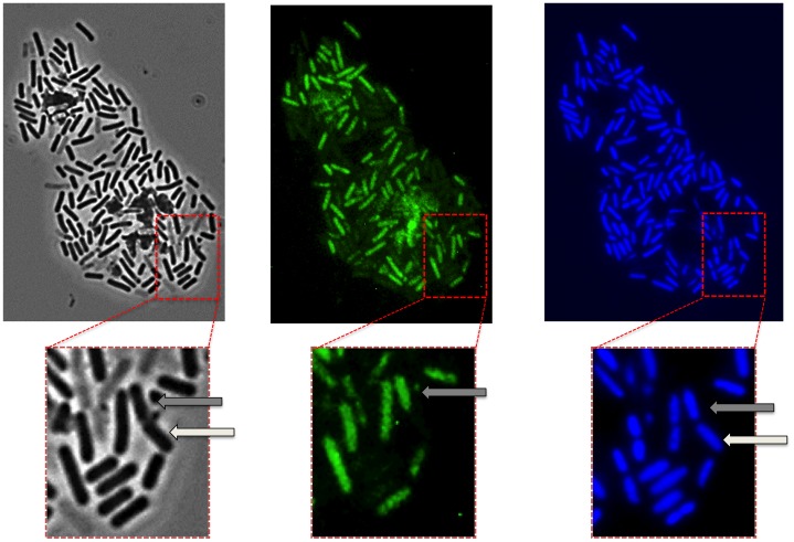
Figure 2. Phase contrast and fluorescence microscopy analysis of SF214.
Observation of the same microscopy field by phase contrast (left), autofluorescence (middle) and DAPI staining (right). The same section of each panel is enlarged. The arrows in the enlarged sections point to a doublet of cells, still partially attached and deriving from the same mother cell, in which only one cell (grey arrows) is autofluorescent.

