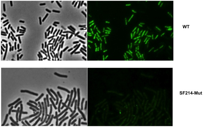
Figure 3. Microscopy analysis of SF214 and of its unpigmented mutant.
Microscopy analysis of SF214 and of its unpigmented mutant (SF214-Mut). For each strain the same microscopy field is shown by phase contrast (left) and autofluorescence (right). The same conditions of exposure were used for the two microscopy fields.

