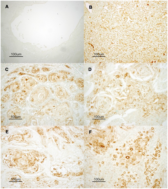
Figure 1. Aberrant expression of AID protein in oral tissues and oral squamous cell carcinoma tissue.
Representative images of immunostaining for endogenous AID are shown. To demonstrate physiological expression of AID protein, normal lingual epithelium (A) and a germinal center in normal neck lymphoid tissue (B) were reacted with an anti-AID antibody. The germinal center containing AID-expressing B lymphocytes shows clear positive staining (brown). However, no AID expression was observed in normal lingual epithelium. Representative moderate-to-strong AID immunostaining is shown in the tumor tissues of oral squamous cell carcinoma classified as T2 (C, D) and neck metastatic tissues classified as N1 (F) and N2 (E). (×200).

