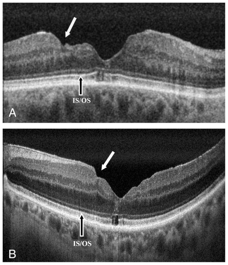
Fig. 3.

High-resolution FD-OCT B-scan images of eyes that had ILM peeling for surgical closure of macular hole. A. An eye with BCVA of 20/60 showing more severe macular surface irregularity (solid white arrow). This eye underwent a second vitrectomy with ILM peeling for recurrent macular hole. Note also the foveal photoreceptor IS-OS abnormality. B. An eye with BCVA of 20/30 with more subtle macular surface irregularity (solid white arrow) and mild epiretinal membrane peripheral to the irregularity. Foveal photoreceptor IS-OS abnormality is present again. Image A is a single frame image and Image B is an average of 10 frames.
