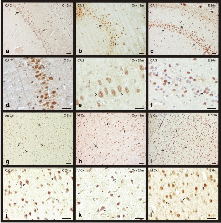
Fig. 3.
Immunohistochemical staining for ERα in the telencephalon of the three experimental groups: control (C), ovariectomized (OVX), and estradiol treated (E) animals during aging (0–24 months). a–f ERα presence in CAs of the hippocampus. g–l ERα presence in some cortical regions: the auditory cortex, motor cortex, visual cortex, and olfactory cortex. Nuclear location of ERα (arrows). Citoplasmatic location of ERα (arrowheads). (bar = 200 μm) (m months)

