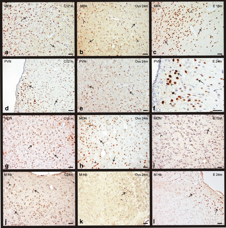
Fig. 7.
Immunohistochemical staining for ERα in the diencephalon of the three experimental groups: control (C), ovariectomized (OVX), and estradiol-treated (E) animals in different age groups (0–24 months). a–c ERα presence in the medial preoptic area (MPA). d–f ERα presence in the paraventricular nucleus (PVN). g–i ERα presence in the medial dorsal nucleus (MDN). j–l ERα presence in the medial habenular nucleus (M Hb). Nuclear location of ERα (arrows). Citoplasmmatic location of ERα (arrowheads). (bar = 200 μm) (m months)

