Figure 2.

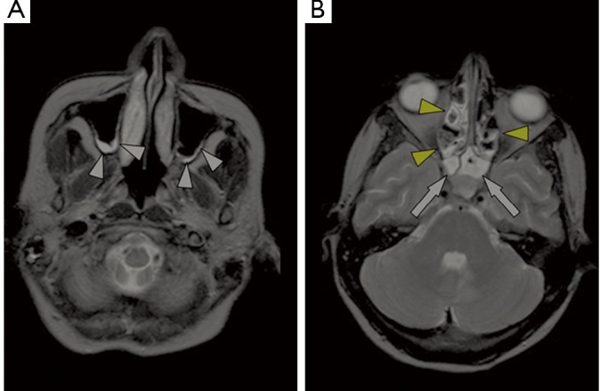
2D axial T2-weighted MRI images of an abnormal patient (corrected LM score=15) shows the hyperintense thickened sinus mucosa in the maxillary (white arrowheads), ethmoid (yellow arrowheads) and sphenoid sinuses (white arrows)

2D axial T2-weighted MRI images of an abnormal patient (corrected LM score=15) shows the hyperintense thickened sinus mucosa in the maxillary (white arrowheads), ethmoid (yellow arrowheads) and sphenoid sinuses (white arrows)