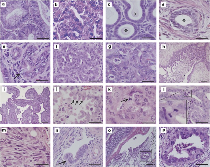
Figure 2.
Pot1a p53 tumors resemble human Type II EMCAs. H&E-stained tissue sections. (a) Human Type I endometrioid adenocarcinoma. (b) Human Type II serous carcinoma. (c) Sprr2f-Cre; Pot1aL/L mouse, histologically normal endometrial glands at 9 months of age (asterisks). (d) Sprr2f-Cre; p53L/L invasive EMCA, uterus. Tumor is composed of well-differentiated invasive glands with relatively minor atypia (asterisk). (e) EIC-like lesion with severe atypia in uterus from 9-month-old Sprr2f-Cre; Pot1aL/L; p53L/L mouse. Arrow points to highly atypical cell with large nucleus; other cells in field display varying degrees of pleomorphism and atypia. (f–p) Representative histologies from different Sprr2f-Cre; Pot1aL/L; p53L/L animals. (f, g) Representative areas of tumor showing high-grade features. (h) Tumor with focal micropapillary features. (i) Tumor with fibrovascular papillary architecture. (j) Hobnailing of tumor cells (arrows). (k) Representative high-grade tumor with tripolar mitosis consistent with severe aneuploidy (arrow). (l) Anaphase bridge consistent with presence of telomeric end-to-end fusions. (m) Focal sarcomatous differentiation, an example of extreme de-differentiation strongly associated with aggressive behavior. (n) Lymphovascular invasion by tumor away from the main tumor mass (arrow). (o) Colon with metastasis to pericolonic fat. (p) Higher magnification of metastatic gland in (o). Scale bars=20 μℳ for (c) and (e), 100 μℳ for (h), (i) and (o) and 50 μℳ for all other panels.

