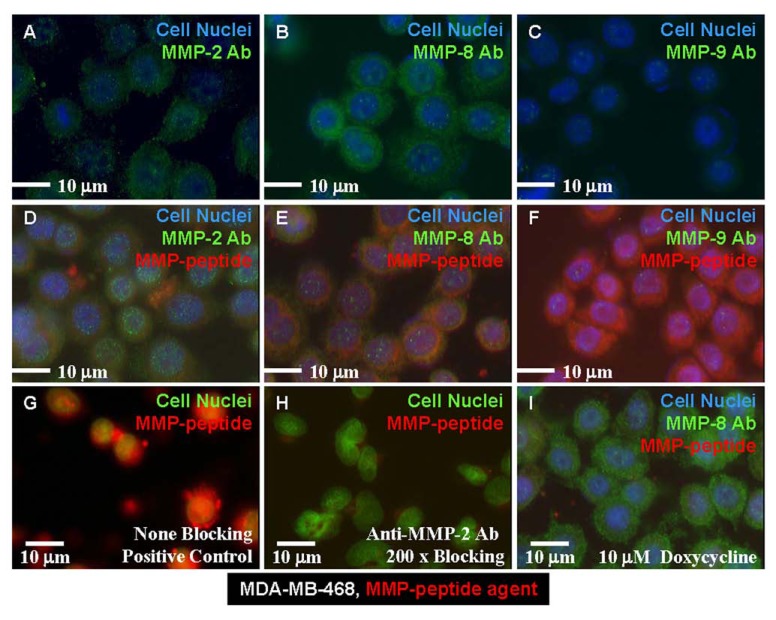
Fig. (2).
MMP cell images. Human breast cancer cell line (MDA-MB-468) was positive for MMP-2 expression (A), strongly positive for MMP-8 (B), and weakly positive for expression of MMP-9 (C). (D) The MMP-peptide agent had the same binding site as did the anti-MMP-2 Ab. (E) The same peptide had a different binding site than that of the anti-MMP-8 Ab. (F) MMP-peptide bound to different motif than MMP-9 Ab. (G) The peptide agent bound to none of the positive control cells that had been treated with blocking Ab. (H) Anti-MMP-2 Ab blocked the peptide binding to the cells. (I) Doxycycline-treated cells lost the capability of binding to the MMP peptide agent, but were still able to bind the anti-MMP-8 Ab.

