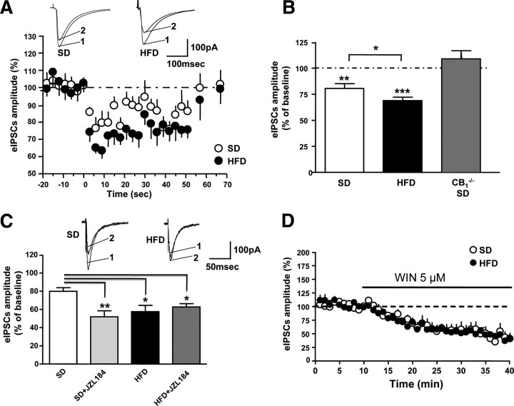
Figure 6.
A, DSI in CA1 hippocampal pyramidal neurons of HFD- and age-matched SD-fed mice. Bottom panel, Normalized eIPSCs amplitude with 3 s depolarization steps (HFD-fed mice: n = 12, filled circles; SD-fed mice: n = 11, open circles). Top panel, Representative traces of eIPSCs before (1) and after (2) DSI induction. B, Summary of DSI magnitude of SD- and HFD-fed wild-type mice, and CB1−/− SD-fed mice. Data are mean ± SEM. **p < 0.001, ***p < 0.01 versus respective baseline before depolarization (100%, dotted line). C, Effect of the inhibitor of 2-AG degradation JZL184, 100 nm on DSI. Bottom panel, Summary of DSI magnitude in SD-fed and HFD-fed mice before and after application of JZL184. Data are mean ± SEM. *p < 0.05, **p < 0.01 versus SD-fed mice without JZL184 treatment. Top panel, Representative traces of eIPSCs, 3 s after depolarization in untreated (1) and JZL184-treated (2) slices. D, Application of the CB1 agonist WIN-55,212-2 (WIN) (5 μm) strongly reduced GABAergic currents with no significant difference between two diet groups.

