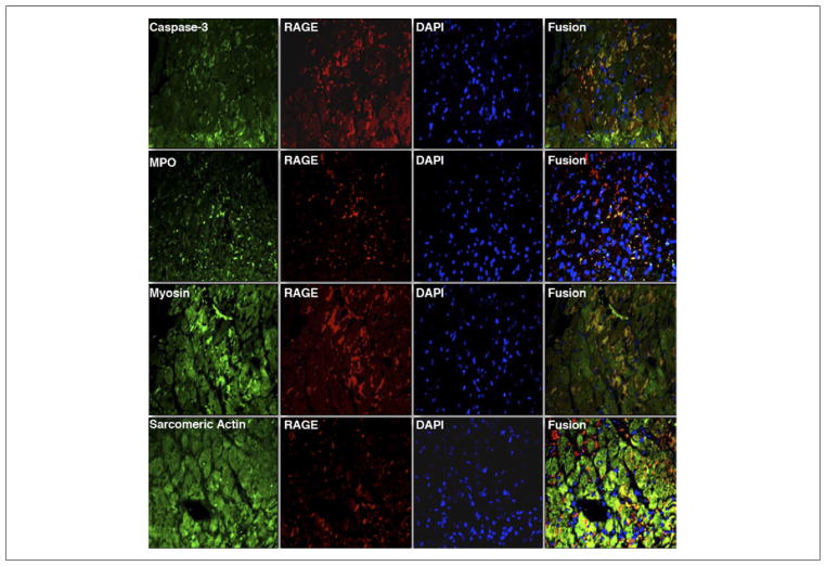
Figure 7. Representative Dual Immunofluorescent Staining for Cells Expressing RAGE in the Infarcted Myocardium.
Serial sections stained for receptor for advanced glycation end products (RAGE) (anti-RAGE, Texas red) were costained with α-active caspase-3 (apoptotic cells, fluorescein isothiocyanate [FITC]), myeloperoxidase (MPO) (leukocytes, FITC), α-sarcomeric actin (cardiomyocytes, FITC), and α-myosin antibody (myocyte injury, FITC). Counterstaining with 4′,6-diamidino-2-phenylindole (DAPI) (blue) was performed to identify nuclei. Areas in yellow in the merged images represent colocalization. (Magnification ×400).

