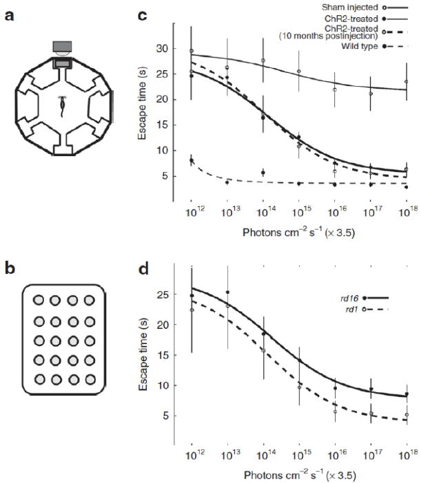
Figure 1. Expression of ChR2-GFP in retinal on bipolar cells leads to improved visual performance.

(a) The schematic for the water maze used to measure perceptual threshold (i.e., the minimum light necessary to guide the escape behavior). The escape platform was tethered to a full spectrum 5 × 4 LED array target (b) with a maximum blue light output of 3.5 × 10¹⁸ photons/cm−2/s as measured at 470 nm. The ambient light of the room was dim and at least two orders of magnitude below the LED array target output. After 14 training sessions, escape time was measured at different light levels (x axis in c and d). We compared the behavioral performance of rd1 and rd16 mice treated with AAV8-Y733F (mGRM6-SV40-ChR2-GFP) (c) relative to the performance of untreated rd1 mice and normally sighted wild-type C57Bl/6 mice. Generally, both the rd1 and rd16 mouse models of blindness had improved escape times as a function of increasing light intensity, however, rd1 mice appear to perform better than rd16 (d). ChR2, human codon-optimized channelrhodopsin-2; GFP, enhanced green fluorescent protein. Adapted from ref. [4].
