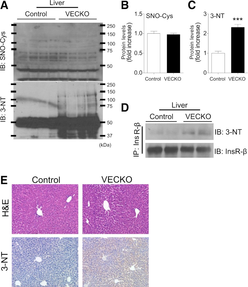
FIG. 5.
InsR nitration in liver. A: Immunoblots (IB) and quantification of S-nitrosylation (SNO-Cys) (B) and nitrotyrosine (3-NT) (C) in liver from 12-week-old WT and VECKO mice (n = 4). D: Immunoblots of nitrotyrosine in liver tissue after immunoprecipitation with InsR. E: Representative photographs of hematoxylin-eosin (H&E) and immunostaining of nitrotyrosine in liver sections. (A high-quality color representation of this figure is available in the online issue.)

