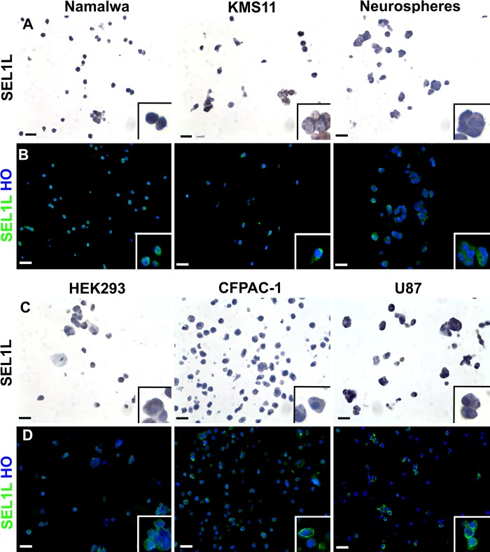
Figure 1.
Immunocharacterization of six cell lines by cell microarray technology. After cell collection, pellets deriving from six different cell lines were resuspended in agarose and then embedded in paraffin. Two consecutive sections were analyzed for SEL1L expression by immunohistochemistry (A, C) and immunofluorescence (B, D) techniques (using the monoclonal antibody described by Orlandi et al. 2003), showing the typical perinuclear/cytoplasmic subcellular location of this protein. In the immunohistochemical images, the brown color reflects SEL1L expression and nuclei were counterstained with hematoxylin; differently, in immunofluorescence analysis, SEL1L is represented in green and nuclei in blue (counterstaining with Hoechst 33258, HO). Scale bars = 10 µm.

