
Figure 1.
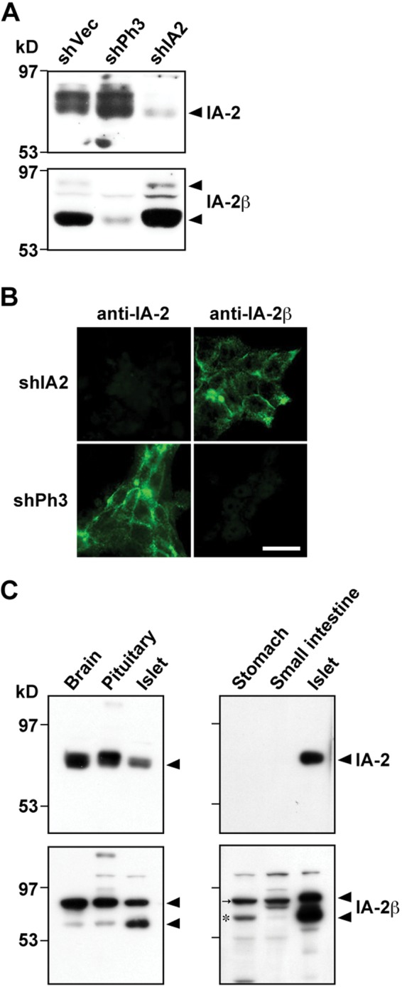
Immunoblot and immunofluorescence analyses with antibodies against islet-associated protein–2 (IA-2) and IA-2β in INS-1 rat pancreatic β-cell line and rat tissues. (A) INS-1 cells were infected with the adenovirus integrating or expressing shVector (shVec), shIA2 (shRNA for IA-2), or shPh3 (shRNA for IA-2β). After 48 hr, cell extracts were prepared, and each extract normalized for total protein content (20 µg) was subjected to immunoblotting with anti–IA-2 (upper panels) or anti–IA-2β (lower panels). (B) The INS-1 cells expressing shIA-2 or shPh3 were fixed and immunostained with antibodies against IA-2 or IA-2β. Bar, 10 µm. (C) Rat tissue lysates (4 µg of protein content extracted from the whole brain, pituitary, and pancreatic islets [left panels]; 100 µg of protein content extracted from stomach and small intestine; and 1 µg of islet protein [right panels]) were subjected to SDS-PAGE/immunoblot analysis. An asterisk indicates specific bands labeled with anti–IA-2β antibody in the stomach and small intestine (very weak), and an arrow indicates nonspecific bands in the same tissues.