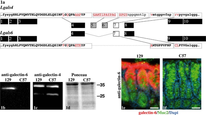
Figure 1.
Anti-galectin-4 and -6 specificity. Organization of the Lgals4 and Lgals6 exons in relation to the structure of the galectin-4 and galectin-6 proteins (1a). The exons encoding the carbohydrate recognition domains of galectin-4 and -6 are shown in black. The exons encoding the linker region of galectin-4 deleted in galectin-6 are shown in grey. The exon 7 that encodes the part of the galectin-4 linker region still present in galectin-6 is shown in white. The sequence of the epitopes used to produce the antibodies directed against galectin-4 and -6 are shown in capitals at the top (galectin-4) and bottom (galectin-6) of the figure. The boxes isolate the sequences from the corresponding exons. The epitope residues conserved between the galectin-4 and -6 proteins are in bold, and those not conserved are in red and underlined. Western blot on colon samples with the anti-galectin-6 (1b) and anti-galectin-4 (1c) antibodies, as well as a Ponceau S red staining to assess the quality of the transfer (1d). The mouse strain from which the samples were obtained is indicated over each lane (129 standing for 129/Sv, and C57 for C57BL/6J). The antibody or the staining procedure that has been used is indicated at the top. The position of the ladder bands is indicated on the right (in kD). By immunofluorescence, the anti-galectin-6 labels the colonic mucosa in the 129/Sv (1e) but not the C57BL/6J (1f) background. The anti-galectin-6 staining is shown in red, anti-Muc2 in green, and DAPI-stained nuclei in blue. Scale bars in 1e and 1f = 40 µm. The same settings and exposure times were used for both pictures.

