Figure 3.
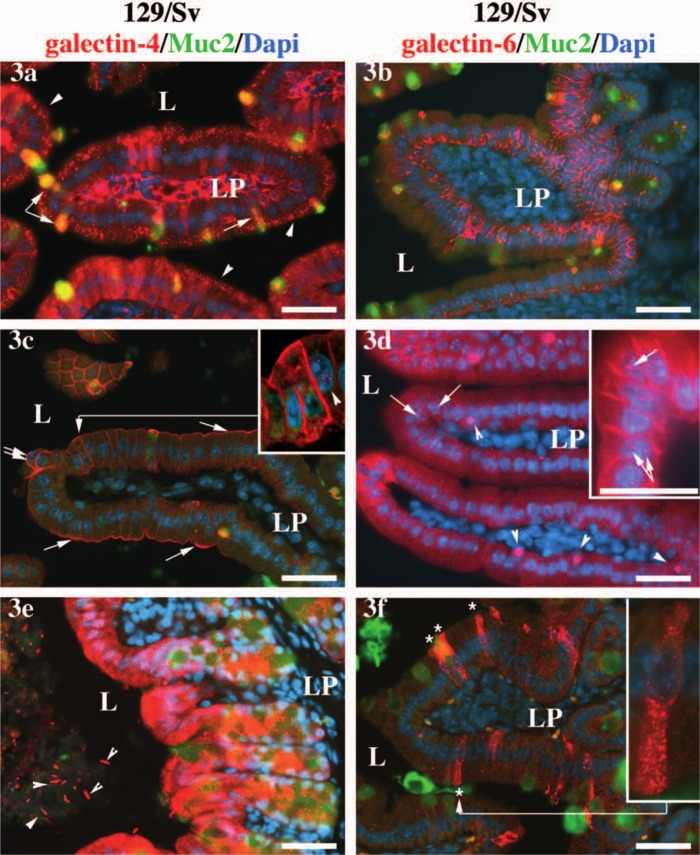
Differences in the subcellular localization of galectin-4 and galectin-6 in the 129/Sv background. Expression of galectin-4 (3a) and galectin-6 (3b) in samples of duodenum fixed by immersion into the fixative, instead of via intracardiac perfusion, as in the rest of the figures. In 3a, the arrowheads point toward endosomes in the enterocytes; the arrow points toward galectin-4 aggregates at the base of the secretion granule of a goblet cell, and the double arrow points toward galectin-4 and Muc-2 positive secretory granules also in goblet cells. In 3c, the arrows point toward an accumulation of galectin-4 protein in enterocyte apical membrane, and the double arrows point toward round shaped cells at the apex of the villus. In 3d, the arrows point toward galectin-6 positive nuclear granules in enterocytes, and the arrowheads point toward large nuclear granules in goblet cells. In 3e, the arrowheads point toward galectin-4-decorated bacteria present in the colonic lumen. In 3f, the asterisks label galectin-6-expressing enteroendocrine cells, and the inset shows an enlargement of one of such cells. The anti-galectin-4 or -6 staining is shown in red, the anti-Muc-2 in green, and the DAPI-stained nuclei is in blue. Scale bars = 40 µm. L = lumen; LP = lamina propria. The same settings and exposure times were used for all pictures. The levels were altered for the inset in 3c in order to enhance the enterocytic nuclear staining (arrowhead).

