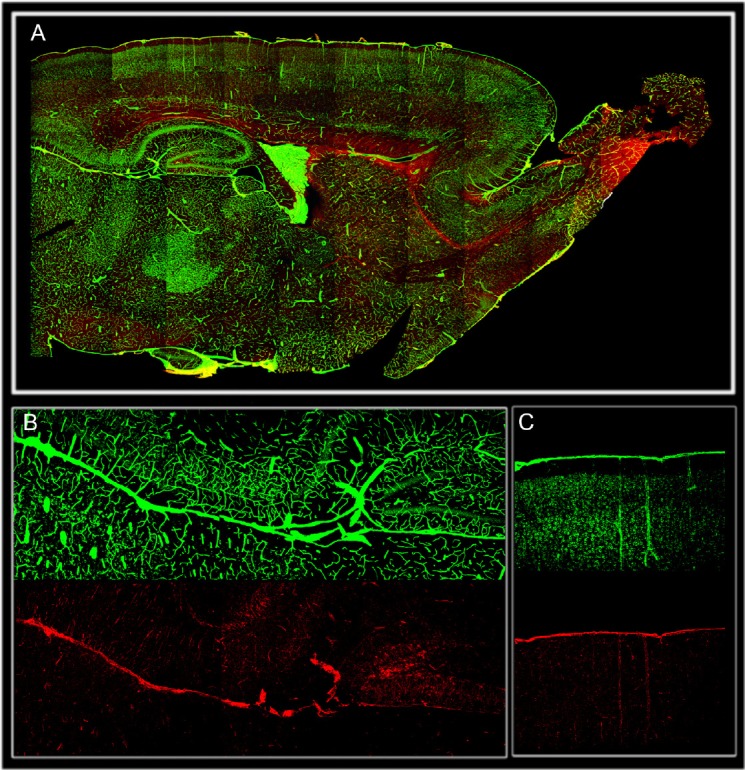
Figure 1.

Distribution of brain meninges (laminin green) and nestin positive cells (red) in 15 days postnatal rat brain transversal section. (A). CNS brain meninges meninges cover and penetrate the brain deeply at every level of its organization including sheaths of blood vessels (perivascular space) and projections located underneath the hippocampal formation that continue with the choroid plexus. High magnification showing nestin positive cells associated with meningeal projection underneath the hippocampus (B), and penetrating the cortex as sheath of blood vessels (C). Meningeal stem cells (nestin) appear to be largely diffuse inside the parenchyma.
