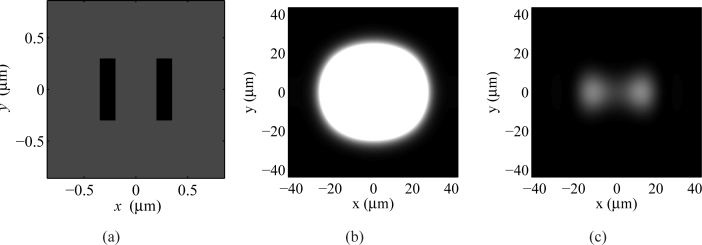
Fig. 8.
Numerical microscope images of two rectangular scatterers buried inside the upper half space, under focused-beam illumination. (a) Refractive index map of the xy cross section at z = 300 nm. (b) The bright-field image of the structure, dominated by the light reflected from the interface. (c) The image with the reflection from the interface removed. This resembles the procedure followed in dark-field microscopy.

