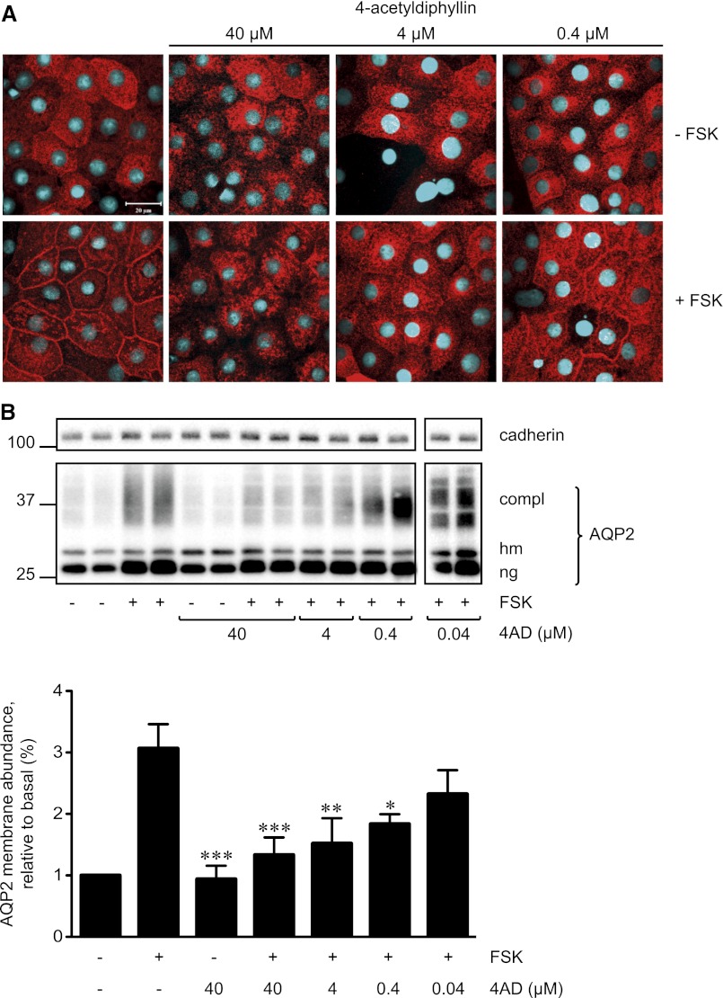
Figure 2.
4AD concentration-dependently inhibits the forskolin (FSK)-induced redistribution of AQP2 in primary IMCD cells. (A) Primary IMCD cells were left untreated and were stimulated with forskolin (10 µM, 20 minutes) or with a combination of 4AD (40, 4, or 0.4 µM, 30 minutes) and forskolin. AQP2 was detected by immunofluorescence microscopy using specific primary antibodies (H27) and Cy3-coupled antirabbit secondary antibodies (red).51 Nuclei were stained with DAPI (blue). Shown are representative images from one of three independent experiments. (B) Upper panel. Cell-surface biotinylation assays were carried out to quantify the effect of 4AD on the localization of AQP2. To detect AQP2 on the surface of IMCD cells, surface proteins were biotinylated with EZ-Link Sulfo-NHS-SS-Biotin, precipitated with streptavidin-agarose, separated by SDS-PAGE, and AQP2, and as a loading control cadherin were detected by Western blotting (shown is a representative blot of three independent experiments). Lower panel. The amounts of complex (compl) glycosylated AQP2 and the high mannose (hm) form of AQP2 and nonglycosylated (ng) AQP2 on the blots were quantitatively analyzed by densitometry; each bar of the histogram represents the total amounts of the three AQP2 forms (mean ± SEM). *P<0.05, **P<0.01, and ***P<0.001 versus forskolin-treated cells.

