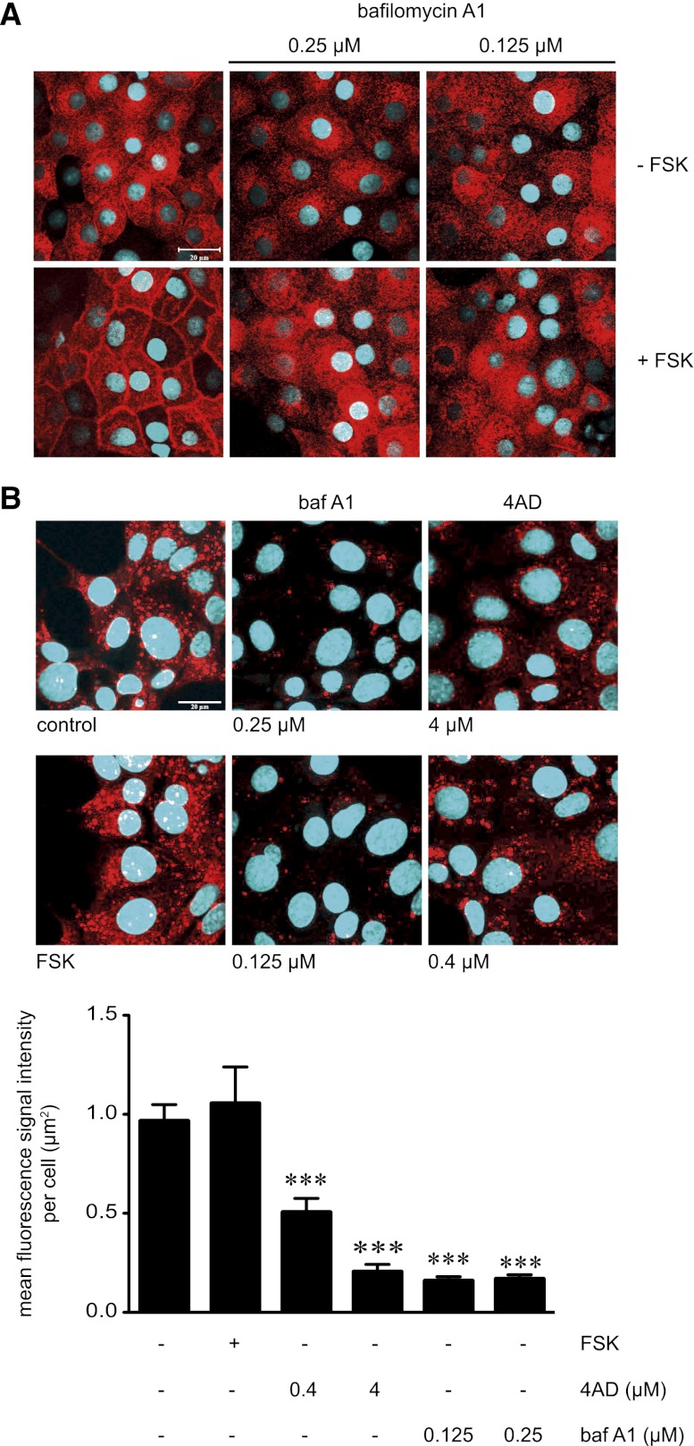
Figure 4.
Inhibition of V-ATPase with 4AD or baf A1 increases acidity of intracellular vesicles in IMCD cells and is associated with inhibition of the FSK-induced redistribution of AQP2 from intracellular vesicles to the plasma membrane. (A) IMCD cells were left untreated or treated with baf A1 in the indicated concentrations (30 minutes) in the absence or presence of forskolin (10 µM, 20 minutes). AQP2 was detected by immunofluorescence microscopy using specific primary antibody H27 and Cy3-coupled antirabbit secondary antibodies (red).51 Nuclei were stained with DAPI (blue). Representative images from one of three independent experiments are shown. (B) Upper panel. To visualize acidic compartments in MCD4 cells in the absence or presence of the V-ATPase inhibitors 4AD or baf A1, the cells were incubated with the acidotropic dye LysoTracker Red DND-99 (75 nM, 30 minutes), which is retained in acidified compartments (red vesicular structures). Nuclei were stained with Hoechst 33258 (blue). Shown are representative images from one of three independent experiments. Lower panel. The intensities of fluorescence signals arising from Lysotracker Red DND-99 were determined in individual cells (>50 cells per condition; three independent experiments; mean ± SEM). Statistically significant differences versus untreated cells are indicated (***P<0.001).

