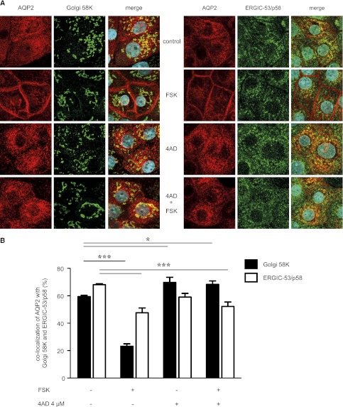
Figure 5.
4AD causes an accumulation of AQP2 in the Golgi compartment. (A) IMCD cells were seeded on 12-mm cover slides, left untreated or treated with 4AD (4 µM or 0.4 µM, 30 minutes). AQP2 was detected with specific primary (C17, Santa Cruz) and secondary Cy2-coupled antibodies (red). ERGIC-53/p58 and Golgi apparatus 58K proteins were detected with specific primary and Cy5-conjugated secondary antibodies (both green). Fluorescence signals were visualized by laser-scanning microscopy. Shown are representative images from one of three independent experiments. (B) The co-localization of AQP2 with ERGIC-53 and Golgi apparatus 58K was determined from the images in part A using ZEN2010 software (Carl Zeiss; >25 individual cells per condition; three independent experiments; mean ±SEM). Statistically significant differences are indicated (*P<0.05; ***P<0.001). FSK, forskolin.

