Figure 6.
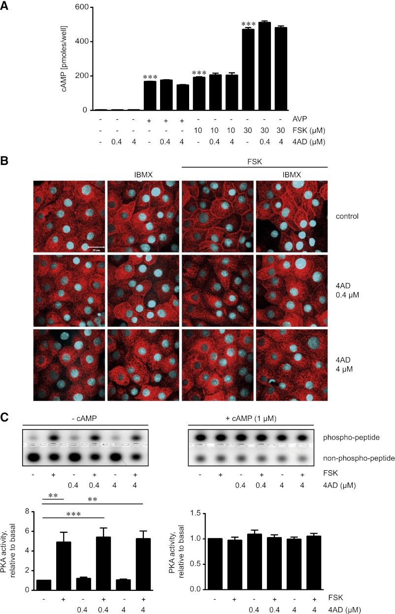
4AD does not affect formation of cAMP or global PKA activity. (A) Primary IMCD cells were left untreated, treated with 4AD (30 minutes), forskolin (FSK; 20 minutes), or AVP (20 minutes) in the indicated concentrations. Where indicated, cells were treated with 4AD for 30 minutes before addition of AVP or forskolin for an additional 20 minutes. Cell lysates were prepared and cAMP concentrations were determined by radioimmunoassay (three independent experiments, triplicates per condition; mean ± SEM; ***P< 0.001). (B) IMCD cells were seeded on 12-mm cover slides, left untreated, or treated with forskolin (10 µM, 20 minutes), IBMX (250 µM, 30 minutes), or the indicated combinations of forskolin and 4AD. AQP2 was detected by immunofluorescence microscopy using specific primary antibody H27 and Cy3-coupled antirabbit secondary antibodies (red).51 Nuclei were stained with DAPI (blue). Shown are representative images from one of three independent experiments. (C) MCD4 cells were left untreated, treated with forskolin (10 µM, 20 minutes), or treated with 4AD in the absence or presence of forskolin as indicated. Upper panels. Cell lysates were prepared and PKA activity was determined by measuring its ability to phosphorylate the substrate peptide PepTag A1. Where indicated, cAMP (1 µM) was added to induce maximal PKA activity. Agarose gels from one of three representative experiments show PKA-phosphorylated and nonphosphorylated PepTag A1 peptide. Lower panels. The amounts of phosphorylated and nonphosphorylated PepTag A1 peptides were densitometrically evaluated. PKA activity is expressed as the ratio of phosphorylated to nonphosphorylated PepTag A1 peptides. Values are mean ± SEM; ***P<0.001.

