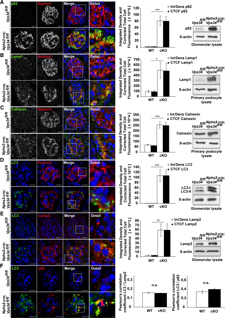
Figure 3.
Autophagy is impaired in Nphs2-cre;Vps34fl/fl mice. Immunofluorescence staining of kidney sections of 3-week-old Nphs2-cre;Vps34fl/fl and littermate control mice. (A–E) Quantification of confocal immunofluorescence microscopy and Western blot analyses of glomerular or primary podocyte protein lysates displayed significant accumulation of the autophagy marker p62, the lysosomal markers Lamp1 and -2, the ER stress marker Calnexin, and the autophagy marker LC3 in podocytes of Nphs2-cre;Vps34fl/fl mice. (E) Confocal microscopy showed no significant colocalization of LC3 and the lysosomal marker Lamp2 in Vps34-deficient podocytes. (F) Colocalization of LC3 and p62 in Vps34-deficient podocytes in confocal microscopy. *P<0.05, **P<0.01, ***P<0.001. Scale bars, 20 µm; 5 µm in detail.

