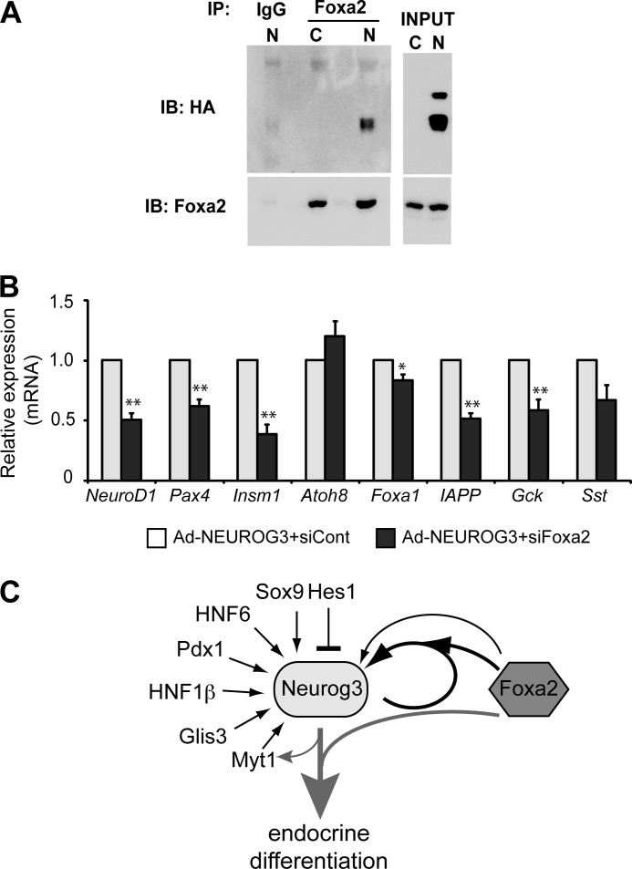
FIGURE 7.
A, mPAC cells were infected with a recombinant adenovirus expressing a HA-tagged NEUROG3 (N) or were left untreated (Control, C). At 48 h after infection, cell lysates were prepared and immunoprecipitated with an anti-Foxa2 antibody or IgG control. Immunoprecipitates were analyzed by immunoblot using goat anti-Foxa2 and mouse anti-HA antibodies. Image is representative of three independent experiments. B, mPAC cells were transfected with a siRNA against Foxa2 (siFoxa2) or control siRNA (siCont). 24 h after transfection, cells were treated with AdCMV.NEUROG3 or left untreated. Total RNA from mPAC cells was collected 44–46 h after infection. Levels of endogenous mRNAs encoding the indicated genes relative to Tbp gene were quantified by real time RT-PCR and expressed as fold-increase versus expression in NEUROG3+siCont-expressing cells, set at 1. Note that all genes tested except for Atoh8 are not expressed in untreated mPAC cells (not shown). All data are presented as the mean ± S.E. from five independent experiments. *, p < 0.05, **, p < 0.01 versus siCont. C, based on our present data we propose a model whereby Neurog3 synergizes with Foxa2 to autoactivate its own expression and to induce the endocrine differentiation program. Foxa2 may also directly regulate the Neurog3 gene. Other known transcriptional regulators of the Neurog3 gene are also depicted.

