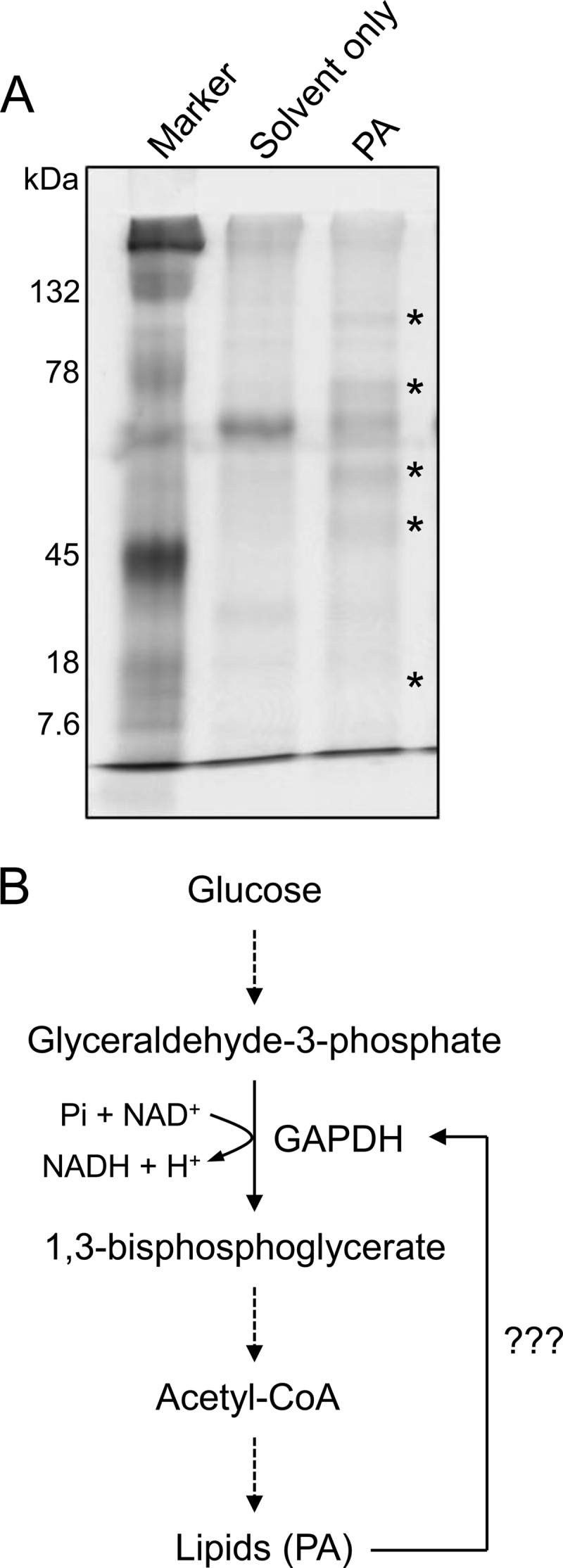
FIGURE 1.

Identification of PA-binding proteins. A, SDS-PAGE image of the candidate PA-binding proteins. Total proteins from C. sativa were incubated with PA-spotted nitrocellulose membrane, and proteins bound to the membrane were resolved by SDS-PAGE. Asterisks indicate the proteins present in the PA-spotted membrane but not in the solvent-only spotted membrane. B, metabolic pathway showing the potential role of GAPDH in lipid biosynthesis. Dashed arrows indicate multistep pathway.
