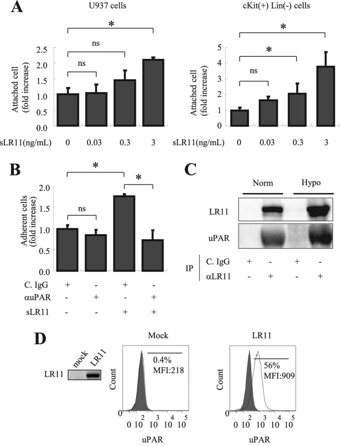
FIGURE 5.

Significance of LR11-uPAR complex formation in hypoxia-induced adhesion of U937 cells. A, U937 cells (left panel) or c-Kit+ Lin− cells (right panel) preincubated for 2 h with the indicated concentrations of sLR11 were subjected to analyses of their adhesion to plates coated with vitronectin as described under “Experimental Procedures.” The numbers of attached cells are presented as -fold increase over those without sLR11 (mean ± S.D. (error bars), n = 3). *, p < 0.05; ns, not significant. B, U937 cells preincubated for 4 h with or without 1 ng/ml sLR11 in the presence of anti-uPAR neutralizing antibody (αuPAR) or of control IgG (C. IgG) were subjected to analyses of their adhesion to plates coated with MSCs as described under “Experimental Procedures.” The numbers of attached cells are presented as -fold increase of those without sLR11 or without antibodies (mean ± S.D., n = 3). *, p < 0.05; ns, not significant. C, U937 cells preincubated for 24 h under normoxic or hypoxic conditions were incubated with 10 ng/ml sLR11 for 15 min, the cell lysates were immunoprecipitated with the monoclonal anti-LR11 antibody M3 (αLR11) or with control IgG (C. IgG) and subjected to immunoblot analysis with the monoclonal anti-LR11 antibody A2-2-3 or with polyclonal antibodies against uPAR, respectively, as described under “Experimental Procedures.” The signals for LR11 (250 kDa) or uPAR (65 kDa) were quantified using the Image Lab software. D, K562 cells stably transfected with cDNA specific for LR11 or vector alone (mock) were subjected to flow cytometric analysis as described under “Experimental Procedures.” The profiles of surface uPAR expression are presented in histograms. The frequencies and the mean fluorescence intensities (MFIs) of the fractions of cells with high uPAR levels are indicated. The filled histograms are isotype controls. Representative data from multiple experiments are shown. Inset, LR11 levels in control or LR11-overexpressing K562 cells were analyzed by immunoblotting using the monoclonal anti-LR11 antibody, A2-2-3.
