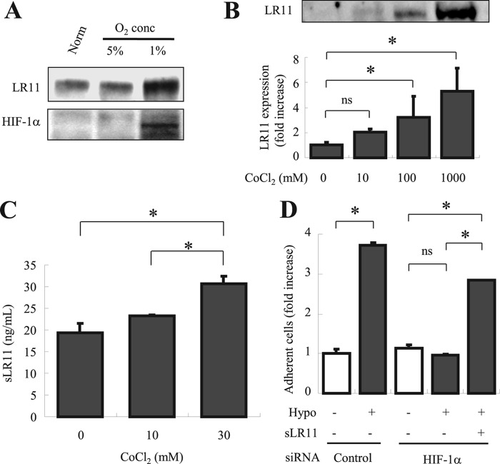
FIGURE 6.
Functional link between HIF-1α and LR11 in enhancing hypoxia-induced cell adhesion. A, after incubation for 24 h under normoxic or hypoxic conditions (1% O2 and 5% CO2), LR11 and HIF-1α protein levels were evaluated by immunoblot analysis with the monoclonal anti-LR11 antibody A2-2-3, or with polyclonal antibodies against HIF-1α, respectively, as described under “Experimental Procedures.” The specific signals for LR11 (250 kDa) and HIF-1α (120 kDa) were quantified using the Image Lab software. B, U937 cells incubated for 48 h with the indicated concentrations of CoCl2 were subjected to immunoblot analysis using monoclonal anti-LR11 antibody A2-2-3 as described under “Experimental Procedures.” The LR11-specific signals at 250 kDa were quantified using the Image Lab software. The blot shown is representative of three independent experiments. Data are presented as mean ± S.D. (error bars; n = 3). *, p < 0.05; ns, not significant. C, conditioned media collected after incubation for 48 h with the indicated concentrations of CoCl2 were concentrated and subjected to sLR11 measurement by ELISA as described under “Experimental Procedures.” Data are presented as mean ± S.D. (n = 3). *, p < 0.05; ns, not significant. D, after incubation in normoxic or hypoxic conditions for 24 h with or without 1 ng/ml sLR11, the numbers of U937 cells previously transfected with siRNA specific for HIF-1α or with control siRNA attached to MSCs were counted. U937 cells incubated for 4 h in the presence or absence of 1 ng/ml sLR11 after preincubation for 24 h under normoxic or hypoxic conditions were subjected to analyses of their adhesion to plates coated with MSCs as described under “Experimental Procedures.” Data are shown as -fold increase compared with control cells in the absence of sLR11 under normoxia. Data are presented as mean ± S.D. (n = 3). *, p < 0.05; ns, not significant.

