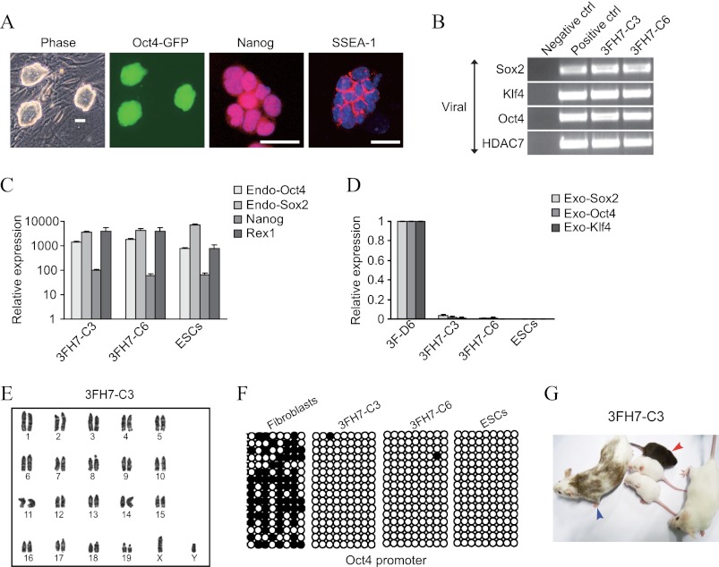
FIGURE 2.
Characterization of iPSCs generated by HDAC7 and OKS. A, phase contrast and immunofluorescence photographs of a representative iPSC clone produced with OKS and HDAC7. Scale bars = 50 μm. B, semiquantitative PCR showing integration of the exogenous transgenes in the genome of selected iPSC clones. Untransduced fibroblasts and pMXs plasmids containing the corresponding cDNA were used as controls (ctrl). C, qPCR for endogenous (Endo) ESC-like markers in the indicated iPSC clones and mouse ESCs compared with fibroblasts. D, qPCR for the exogenous (Exo) transgenes in the indicated iPSC clones. ESCs and reprogramming fibroblasts extracted at day 6 were the controls. E, normal karyotype of a selected iPSC clone generated with OKS and HDAC7. F, DNA methylation status of the Oct4 proximal promoter in the indicated cell types. ○ represent demethylated 5′-cytosine-phosphodiester-guanine sites. G, photograph of a representative chimeric mouse (blue arrowhead) generated with the same iPSC clone. Transmission to the germ line can be observed in the baby mouse with agouti skin color (red arrowhead).

