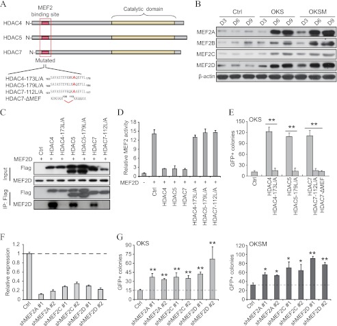
FIGURE 6.
MEF2 proteins are downstream targets of class IIa HDACs during reprogramming. A, schematic depicting the structure of HDAC4, -5, and -7, the location of the MEF2 binding site and the catalytic domain, and the corresponding modifications were performed to abolish their function. B, representative Western blot analysis for MEF2 proteins using lysates of a time course reprogramming experiment with OKS/OKSM in fibroblasts. An empty vector was used as a control (Ctrl, also in D, E, and G). D, day. C, immunoprecipitation of MEF2D after coexpression with the indicated FLAG-tagged class IIa HDAC variants in HEK293T cells. IP, immunoprecipitated samples; Ctrl, control. D, cotransfection of a MEF2 luciferase reporter gene and the indicated expression vectors in HEK293T cells. Activity was measured 48 h post-transfection. Items were measured in triplicate, and the mean values ± S.D. of a representative experiment are shown. E, number of GFP+ colonies in fibroblasts reprogrammed with OKS and wild-type or mutated forms of HDAC4, -5, and -7. F, shRNA vectors for MEF2 proteins were infected in fibroblasts, and the knockdown efficiency was measured by qPCR. Double asterisks indicate p value < 0.01 (also in G). G, number of GFP+ colonies in fibroblasts reprogrammed with OKS/OKSM and the indicated shRNA vectors. Single asterisk indicates p value < 0.05.

