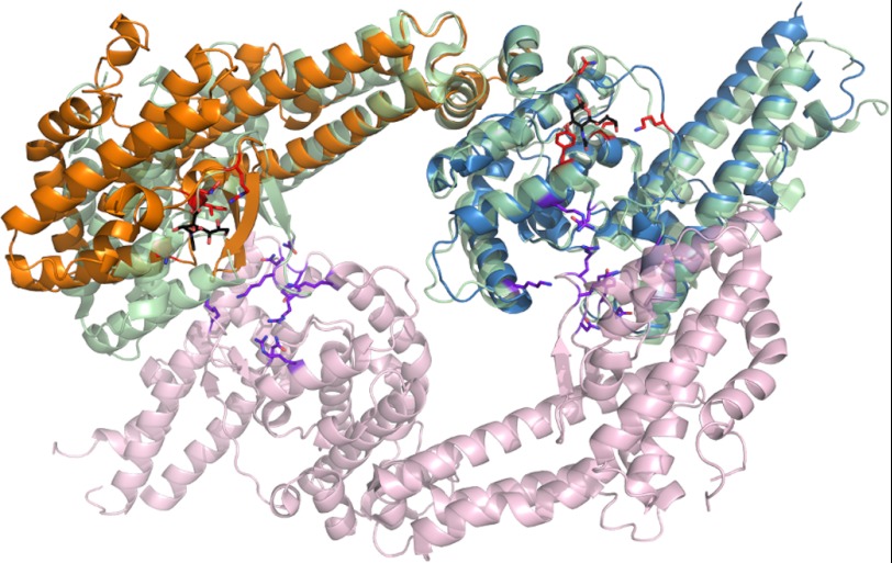
FIGURE 6.
PfEBA-140 sialic acid recognition is distinct from that of PfEBA-175. Shown here is an overlay of the RII PfEBA-140 monomer with the dimer of RII PfEBA-175. The PfEBA-140 F1 domain is shown in orange, and the F2 domain is shown in blue. The RII PfEBA-175 monomer overlaid with RII PfEBA-140 is shown in light green. The second monomer of RII PfEBA-175 is shown in light purple. Residues of PfEBA-140 involved in sialic acid binding are shown in red. The sialic acid molecules bound to RII PfEBA-140 are shown in black for clarity. Residues involved in PfEBA-175 sialic acid binding are shown in purple.

