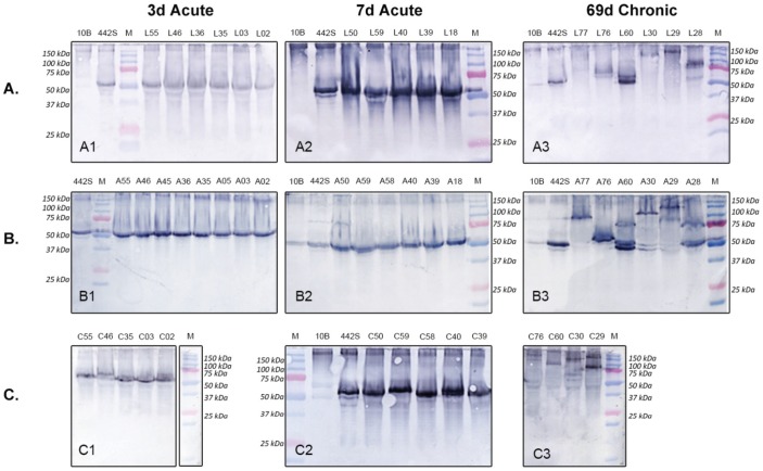
Figure 4. P1 Western blot demonstrating MBA antigenic variation.
Western blots comparing antigenic variation of P1 ureaplasma isolates from animals colonized/infected with U. parvum serovar 3 for 69 days (chronic infection), 7 days and 3 days (acute infection). The number of antigenic variants (single bands) within A. FL (L samples), B. AF (A samples), and C. chorioamnion (C samples) P1 cultures are compared. 442S = serovar 3 initial inoculum control; 10B = 10B media negative control; M = Precision Plus Dual Colour Protein Standard (BioRad, Gladesville, NSW).

