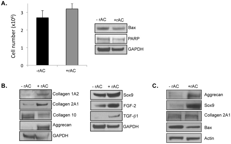
Figure 2. Rat articular chondrocytes were obtained from femurs and grown for 3 weeks in monolayer cultures using standard culture medium with or without recombinant human AC (rAC 200 U/ml).
rAC was added once at the initiation of the cultures. At the end of the 3-week expansion period, the cells were harvested and analyzed. A) Total cell counts (left) revealed no differences in the presence of rAC. Western blotting for two apoptosis markers (Bax and PARP) similarly revealed no differences. B) The expanded chondrocytes were analyzed by western blots for several important chondrogenic markers, including collagens 1A2 and 2A1, aggrecan, Sox9, FGF2, and TGF-beta1. Note that in all cases these chondrocyte markers were elevated in the cells treated with rAC. In contrast to collagens 2A1 and 1A2, collagen 10 expression, a marker of hypertrophy, was lowered by rAC treatment. C) Horse articular chondrocytes were obtained surgically from femoral heads and frozen. The frozen cells were then recovered and grown in monolayer cultures for 3 weeks without rAC. At 3 weeks the cells were passaged and re-plated at a density of 1×10⁶/cm2, and then grown for an additional 1 week with or without rAC. At the end of this 1-week growth period the cells were analyzed by western blots, revealing that the expression of two chondrogenic markers, aggrecan and Sox9, were highly elevated in the rAC-treated horse cells. Bax expression also was reduced in the rAC cells, suggesting a reduction in apoptosis by rAC treatment. All experiments have been repeated at least 3×. Images are representative from individual experiments.

