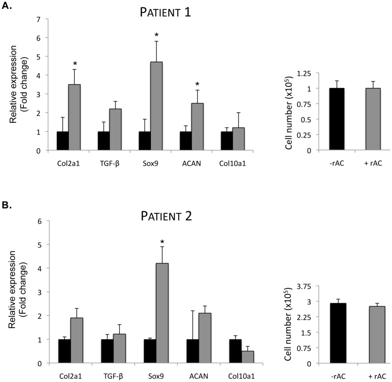
Figure 4. Primary human chondrocytes obtained from the femoral head of a 74-year-old woman with OA were expanded for 3 weeks in monolayer culture with or without rAC added to the culture media (DMEM +10% FBS, rAC added once at the initiation of the culture; 170 U/ml AC).
A) mRNA expression of several chondrogenic markers (collagen 2A1 [Col2a1], TGF-beta1 [TGF], Sox9 and aggrecan [ACAN]) were analyzed by quantitative RT-PCR. Note the positive influence of rAC on the expression of these markers. The expression of collagen 10 was unchanged in these cultures. B) The same experiment was repeated on a second set of cells from a patient with OA. RT-PCR analysis was performed 3×. Consistent with the results obtained with rat and equine chondrocytes (Fig. 2), no significant differences in the number of cells could be found (right panels). * = p<0.05.

