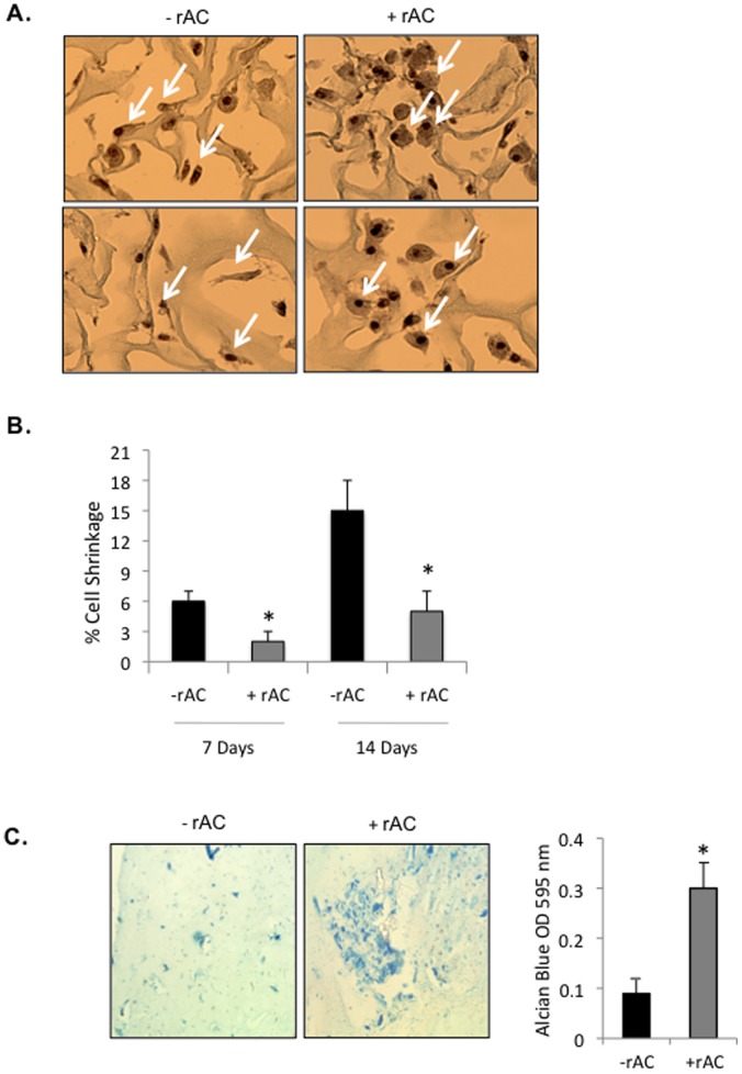
Figure 5. Primary rat chondrocytes were seeded into 3-D collagen scaffolds and grown for 7 or 14 days with or without rAC (DMEM containing 10% serum).
A) They were then analyzed for morphology and proteoglycan production by Safranin-O staining. Note that the cells grown with rAC were larger and maintained a round phenotype that stained positive with Safranin-O (arrows). Shown are representative images from experiments performed 3×. B) Cell shrinkage was evaluated based on Safranin-O and H&E staining using the following equation: DNA unit size = π(fiber segment diameter/2)2 × (myofiber segment length)/myofiber nuclei]. * = p<0.05. C) Primary rat chondrocytes were grown for 2 weeks in biodegradable fibrin gels with or without rAC in the culture media. Alcian Blue staining, another marker of proteoglycan expression, indicated enhanced chondrogenesis (more intense blue color). Experiment was performed 2×.

