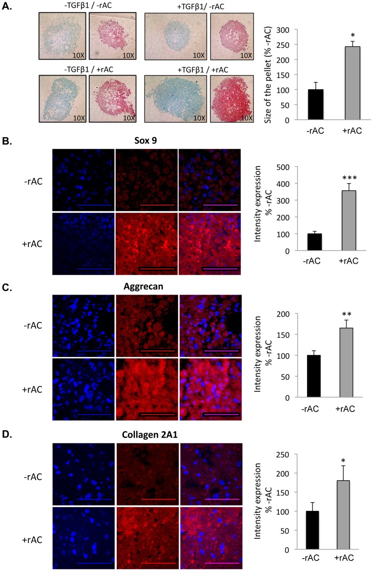
Figure 7. Rat bone marrow cells were isolated and expanded for 3 weeks in standard culture media (without rAC) to prepare homogenous populations of MSCs.
They were then placed into chondrocyte differentiation media (Stem Cell Technology) with or without TGF-beta1 and/or rAC. Pellet cultures were grown for 3 weeks to prepare chondrocytes, and then fixed and analyzed by Alcian Blue and Safranin-O staining, markers of chondrogenesis. A) Note the small and poorly formed pellets in the absence of TGF-beta1 and rAC (upper left). TGF-beta1 is a standard supplement used to induce the differentiation of bone marrow MSCs to chondrocytes. Addition of TGF-beta1 or rAC to the cultures independently had only a modest effect on the pellet size and staining (upper right and lower left). However, inclusion of both proteins in the culture media had a much more significant effect (lower right), both on the size of the pellets and staining intensity. Pellet size is an indicator of the number and/or size of chondrocytes, and staining intensity is a measure of proteoglycan deposition. Beside Alcian Blue and Safranin-O staining, pellets were subjected to immunostaining against B) Sox9, C) Aggrecan, and D) Collagen 2A1. Experiments have been performed with 3 independent rats. Representative images are shown from one experiment. Blue (DAPI) indicates nuclei, and red indicates Sox9, Aggrecan or Collagen 2A1, respectively. Merged images are to the right. Scale bars: 50 µm. * = p<0.05, ** = p<0.005, *** = p<0.001.

