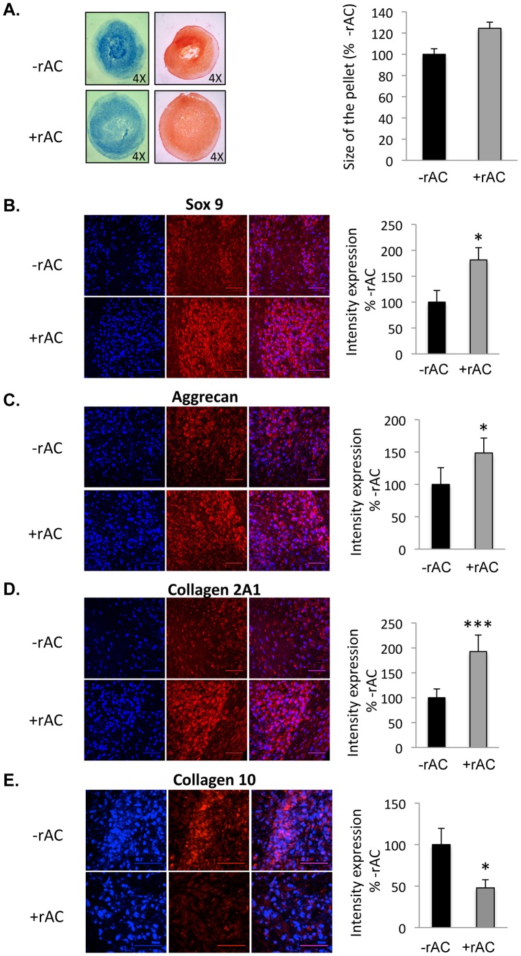
Figure 8. Horse bone marrow cells were isolated and expanded for 3 weeks in standard culture media (without rAC) to prepare homogenous populations of MSCs.
They were then placed into chondrocyte differentiation media containing TGF-beta1, but with or without rAC. A) Pellet cultures were grown for 3 weeks to prepare chondrocytes, and then fixed and analyzed by Alcian Blue (left panel) and Safranin-O (right panel) staining. Note the smaller, more diffuse pellets in the absence of rAC. Pellets were also submitted to immunostaining against B) Sox9, C) Aggrecan, or D) Collagen 2A1. Note the higher expression intensity of Sox9, Aggrecan and Collagen 2A1 in the pellets. E) Pellets were also subjected to an immunostaining against Collagen 10. Note the diminished expression of Collagen 10 in pellets exposed to rAC. DAPI (blue left panel) indicates nuclei, and the right panel (red) indicates Sox9, Aggrecan, Collagen 2A1, or Collagen 10, respectively. Merged images are to the right. Scale bars: 50 µm. * = p<0.05, ** = p<0.005, *** = p<0.001.

